

You are now on the AHDB Horticulture and Potatoes website.
Please click here to access the main AHDB website and other sectors.
Please click here to access the main AHDB website and other sectors.
Go back to the bacterial blotch main page
Brown blotch is frequently caused by Pseudomonas tolaasii (P.tolaasii) and is characterised by dark brown discolouration or spots on the cap surface that can eventually appear as sunken lesions (Figure 1).
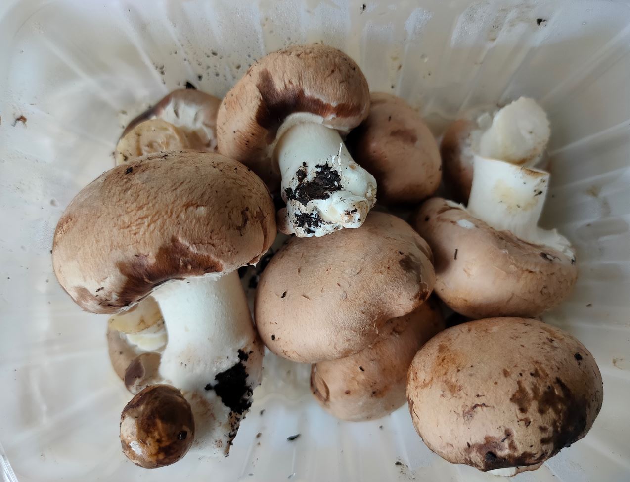
Differences in severity and colour of brown blotch can be dependent on the strain of P. tolaasii. On brown strains of mushrooms, brown blotch can appear as dark discolouration on the underside of caps (Figure 2).
 Ralph Noble
Figure 1: Dark brown discolouration and spot symptoms of brown blotch, caused by a P. tolaassi strain.
 Joana Vicente
Figure 2: Dark discolouration symptoms of brown blotch seen on brown strains of mushrooms.
Other groups of strains in the P. fluorescens species complex, collectively but invalidly named as ‘Pseudomonas gingeri’, cause ginger blotch symptoms. There are at least five different groups of Pseudomonas isolates included in ‘P. gingeri’ that can cause mild to strong ginger blotch in UK farms.
Ginger blotch symptoms typically have a lighter, ginger-coloured appearance, with lesions showing no or only mild sunken appearance (Figure 3). Developed infections of ginger blotch can also causes mushroom cap cracking (Figure 4).
 Ralph noble
Figure 3: Light, ginger discolouration symptoms of ginger blotch, characteristic of a strain of P. fluorescens.
 Ralph Noble
Figure 4: Mushroom cap cracking symptoms of advanced ginger blotch caused by a strain of P. fluorescens
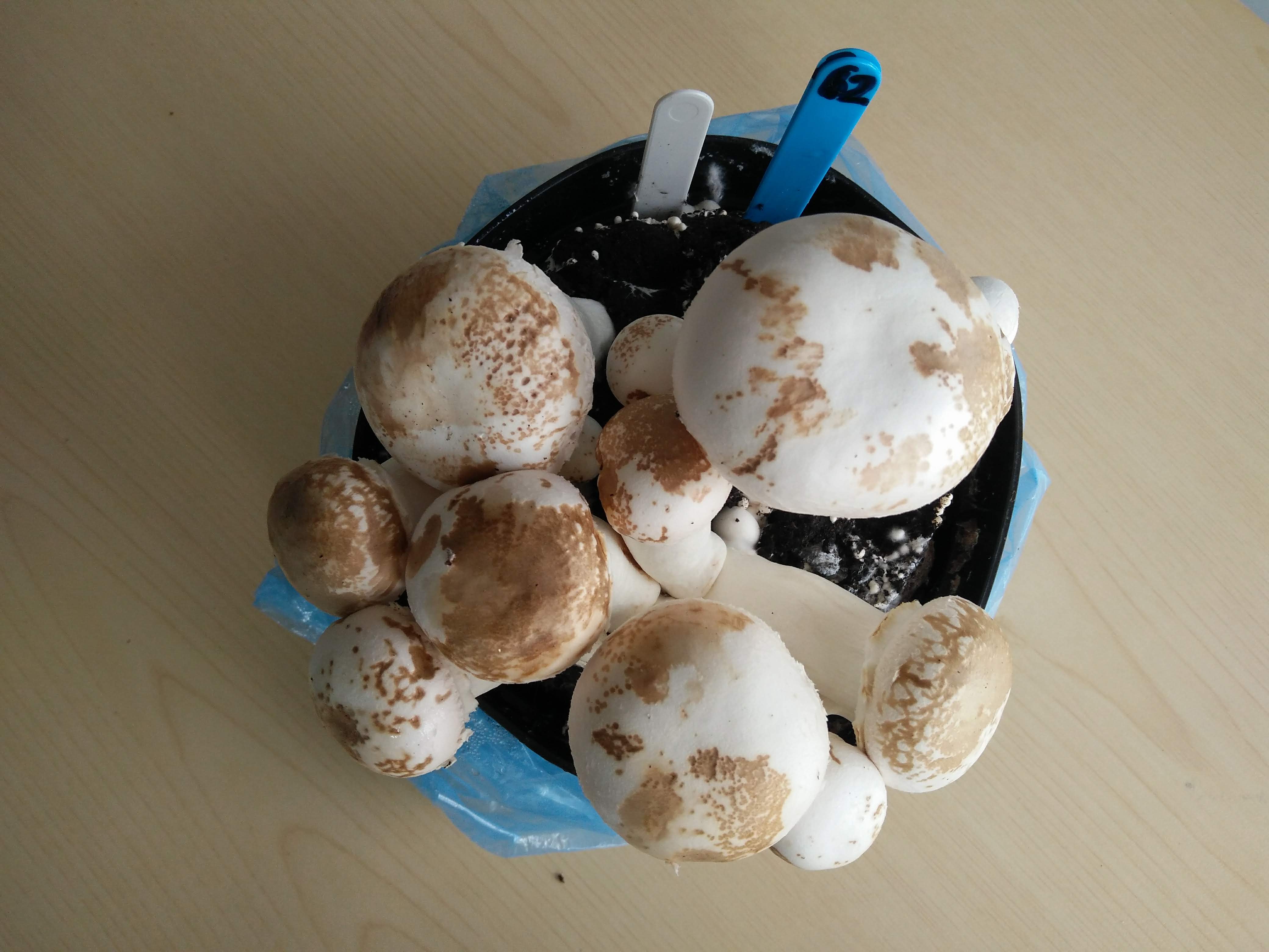
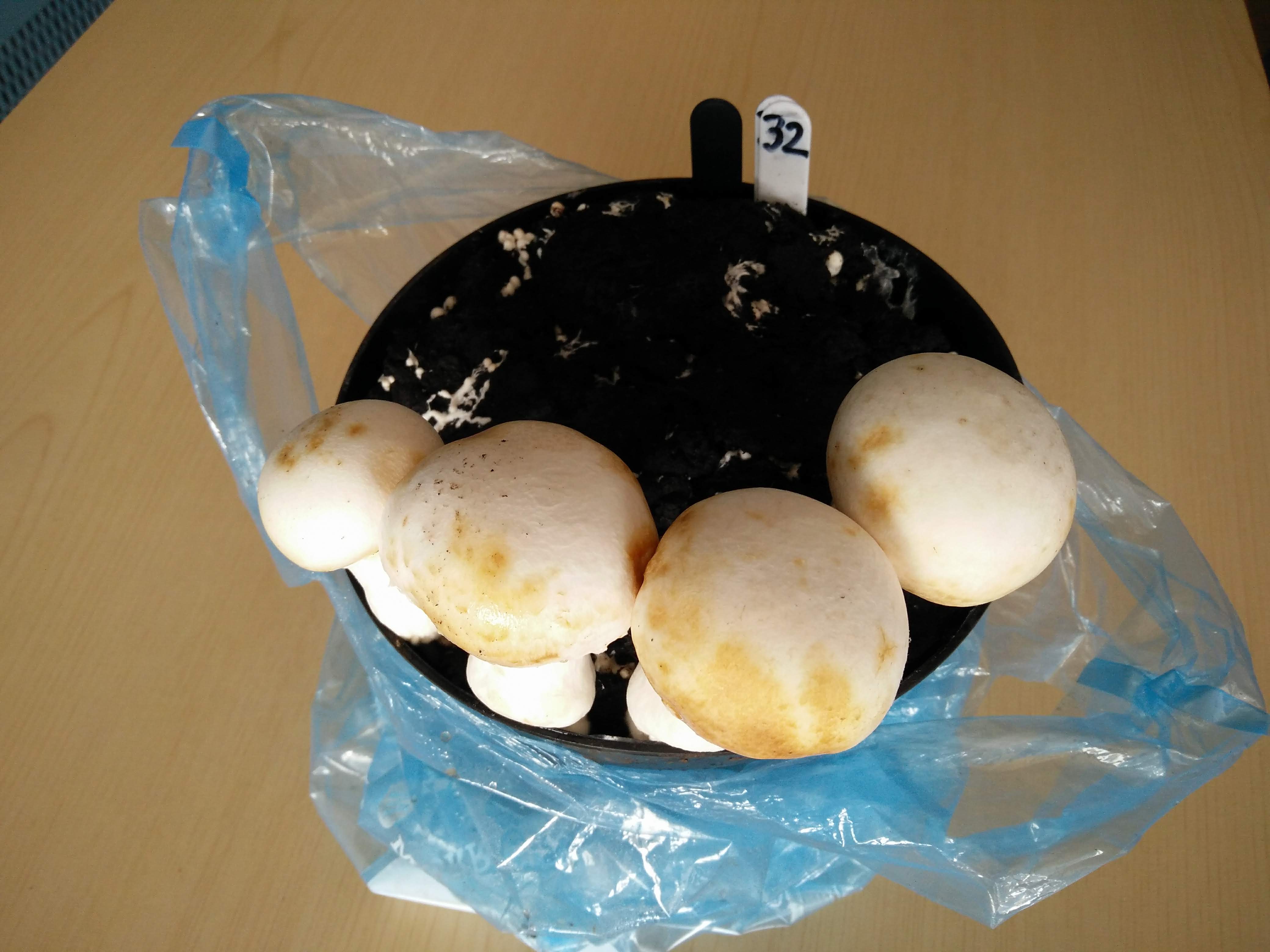
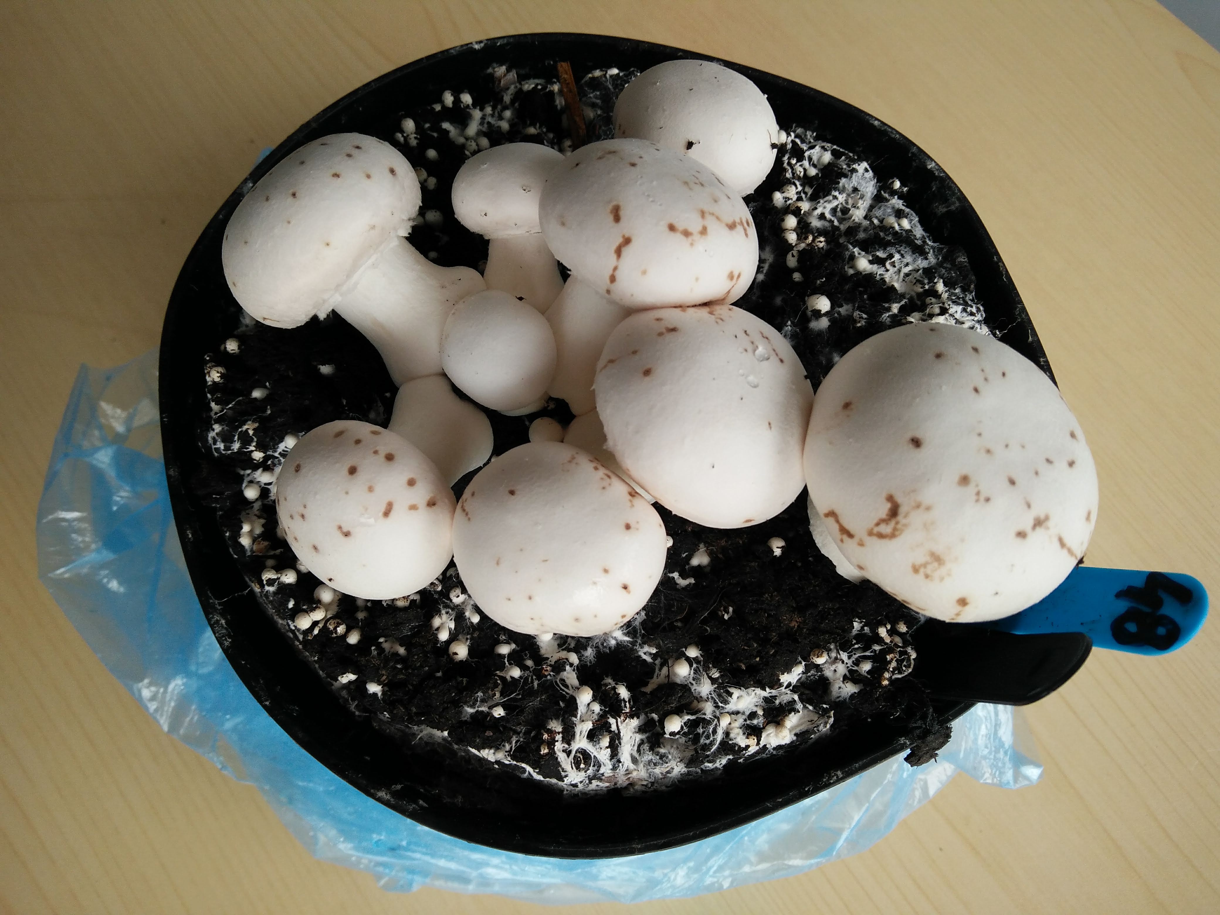
Pseudomonas costantinii causes speckling or pitting on mushroom caps, and brown discolouration, often where mushrooms are touching each other (Figures 5, 6 and 7).
Several other Pseudomonad species such as P. ‘reactans’ and P. agarici can cause blotch symptoms but these are usually mild. P. agarici can cause yellow blotch disease on oyster mushrooms.
 Ralph Noble
Figure 5: Speckling symptoms caused by Pseudomonas costantinii
 Ralph Noble
Figure 6: Pitting symptoms caused by P. costantinii
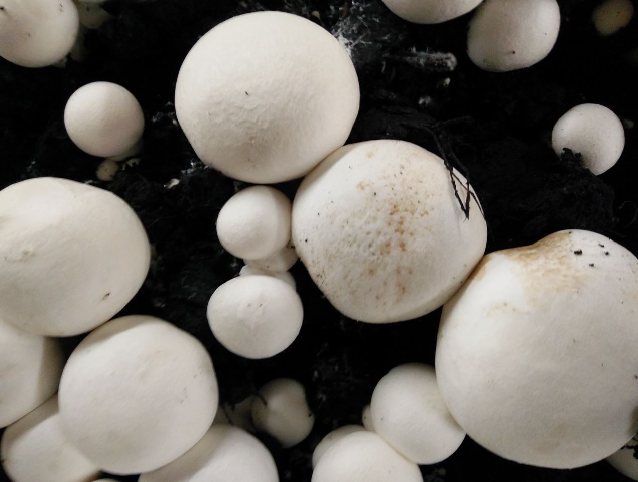 Ralph Noble
Figure 7: Blotch discolouration on caps where mushrooms have been touching caused by P. costantinii
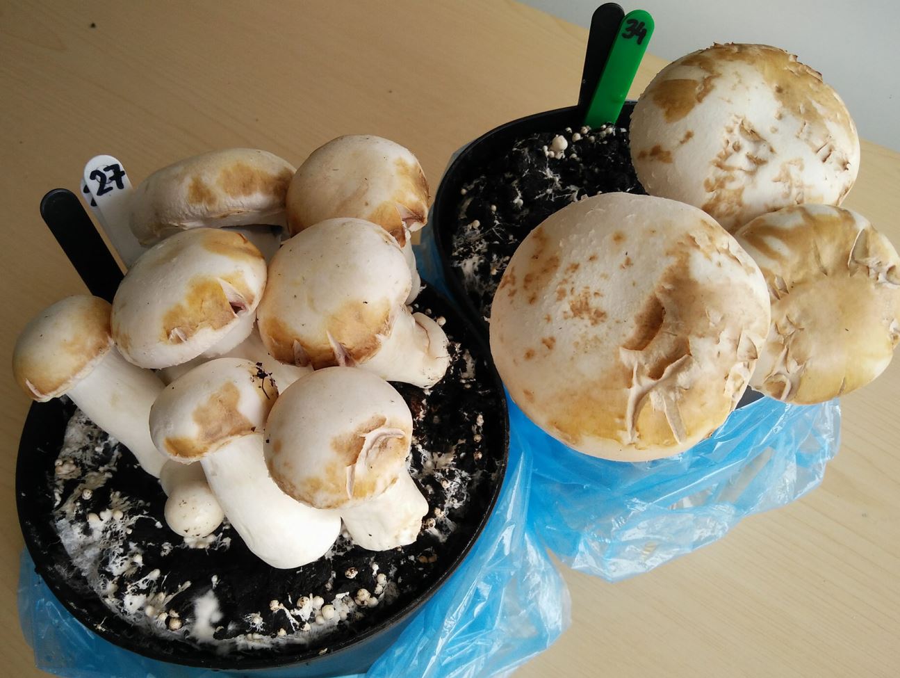
Blotch symptoms vary according to conditions so it can be difficult to diagnose a causal pathogen based on symptoms. Various methods have been developed for testing the blotch pathogenicity of Pseudomonads. Simple mushroom cap tests (Figure 8) can give an indication of blotch pathogenicity especially when disease is caused by P. tolaasii or P. costantinii, but can be unreliable in predicting blotch disease severity in culture tests as seen in Figures 1 to 7.
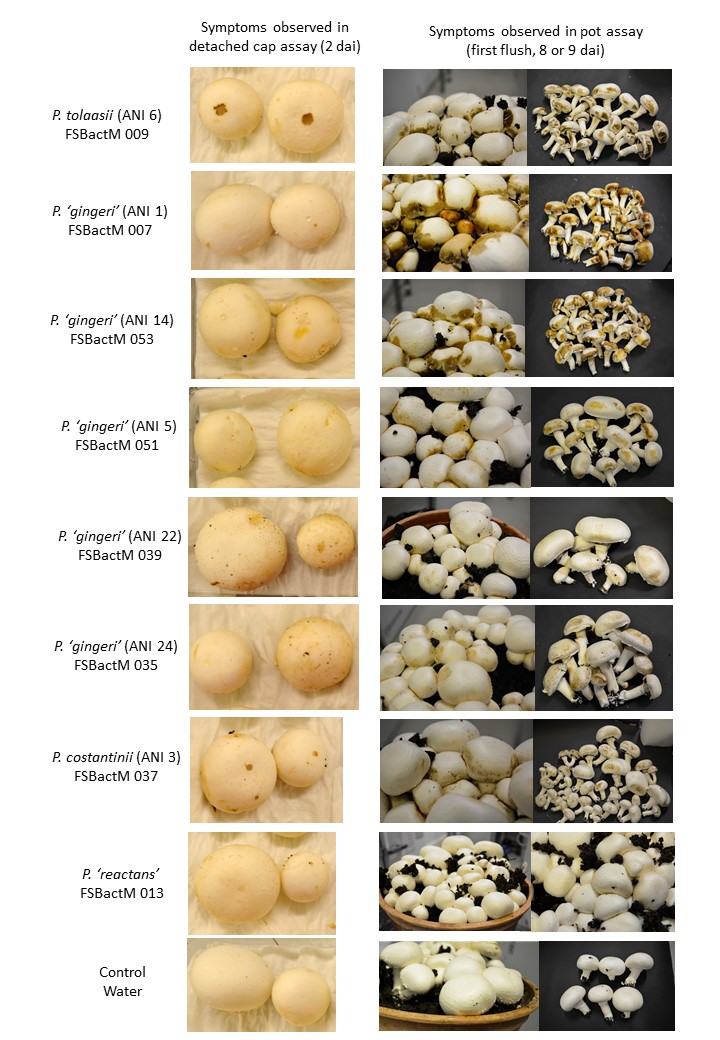
Figure 8: Mushroom cap test vs pot test: Symptoms of blotch caused by different Pseudomonas species in cap and pot assays
Trichoderma aggressivum in mushrooms
Dr Joana Vicente (Fera Science Ltd), Dr Ralph Noble (Microbiotech Ltd) and Professor George Salmond (University of Cambridge)